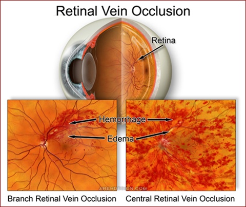
This entity is basically a partial vein occlusion, yet the visual prognosis is better than a central retinal vein occlusion (CRVO). Only a portion or “branch” of the retinal vascular “tree” becomes involved. There is also a much lower incidence of neovascular glaucoma related to the reduced amount of retinal ischemia. Vision is reduced if the functional center of the retina, the macula, becomes involved. If the macula is not involved, central vision may be normal. At times, patients may be completely asymptomatic!
Macular edema, or swelling of the macula, results from the adjacent vein becoming occluded. The goal of treatment is to reduce the amount of macular edema with either laser photocoagulation or intravitreal injection of either steroids or a VEGF inhibitor such as Avastin®, Macugen® or Lucentis®. There was a time when surgery was attempted, but it has not been too popular for a while (this usually means results are not conclusive, i.e. the procedure did not work that well). The goal of any treatment, however, is to minimize the amount of macular edema to yield the best possible vision.
The mechanism by which branch retinal vein occlusions occur involves compression of the adjacent vein as it crosses a retinal artery. At these junctions, there exists a tight ring of connective tissue which surrounds both artery and vein. A hardened, or arteriosclerotic, artery may compress the softer underlying vein. At this point, fluid and blood may leak out into the surrounding tissue causing edema.
