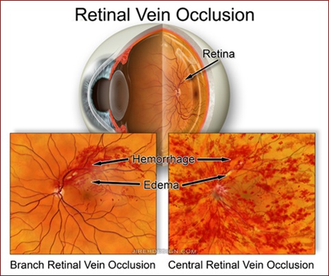
Central retinal vein occlusions may occur in perfectly normal individuals. While there is a higher incidence in patients with hypertension and diabetes, the majority of patients are considered systemically healthy.
In essence, the major retinal vein suddenly closes or becomes severely constricted resulting in loss of blood flow. The result is a very ischemic retina which is accompanied by sudden, painless loss of vision.
In general, there is NOTHING to do to restore vision. Vision loss is usually permanent, however, things can get worse. This is a common mechanism by which a very a painful type of glaucoma may develop. Neovascular glaucoma may result when abnormal blood vessels develop and grow, as a direct result of ischemia, and cover the drainage portions of the eye (the so-called “angle”). The internal fluid, or aqueous humor, can not properly leave the eye and the pressure in the eye sky-rockets. This can lead to severe pain and complete loss of vision.

While there is generally nothing to do to improve vision, the stages of neovascular glaucoma can usually be avoided with frequent, routine follow up. If neovascularization is noted, panretinal photocoagulation (PRP) may be performed to arrest this phase of the disease.
Attempts to restore blood flow, and thus the retinal ischemia, have been of fair success. Radial optic neurotomy has been performed with moderate success. Reports of improvement vary, but are promising. It’s affect on neovascular glaucoma is not proven.
Intravitreal triamcinolone (steroids) and anti-VEGF injections may also be helpful.